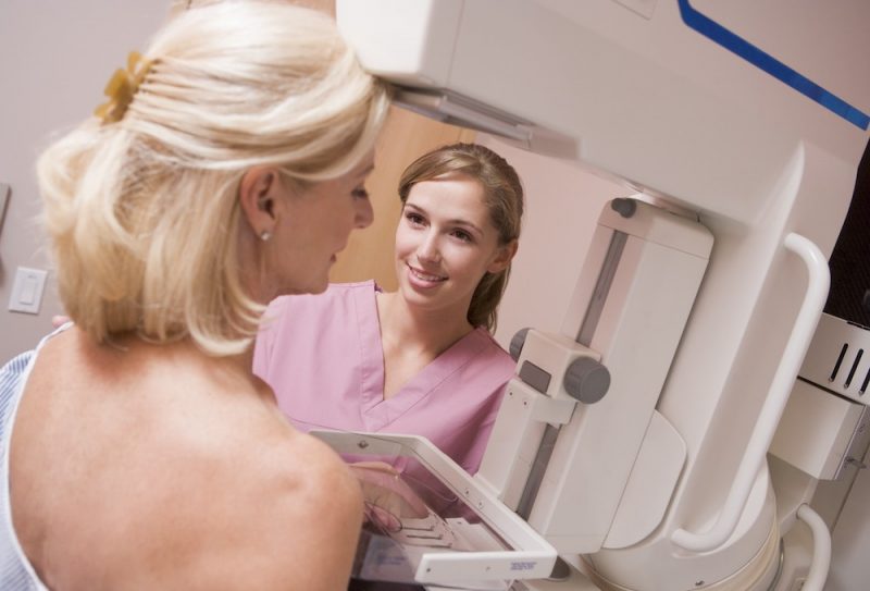
Women with a slower decrease in breast density over time are at higher risk of developing breast cancer. The prospective cohort study, published in JAMA Oncology, online April 27, suggests that dynamic changes in breast density over time can be used to refine risk stratification and guide more individualised screening.
“By adding the change in density over repeated images to models for risk classification in each breast, we set the stage for a better risk estimation with each updated mammogram. We can then better classify the future risk and refer women to appropriate prevention strategies such as enhanced screening as part of routine breast health services,” says senior author Graham Colditz, an epidemiologist from Washington University School of Medicine, St Louis, Missouri.
Mammographic breast density is a well-established risk factor for breast cancer. One-time assessment of breast density represents a key component for risk stratification, with a systematic review demonstrating that the addition of a one-time density measurement increased discriminatory accuracy in seven of 11 studies. As a result of such studies, volumetric density has been incorporated into existing risk assessment models, including the Tyrer-Cuzick model version 8. Traditional models for risk prediction focus on information gathered at baseline, with limited data on the association between breast density changes with time and the risk of subsequent breast cancer diagnosis.
Colditz and colleagues viewed repeated mammograms as an untapped source of data on breast density and hypothesised that individual breast changes over time might shed more light on the relationship between breast density and disease. “A premalignant breast lesion must develop, grow, and become sufficiently large to be detected. Hence one can postulate that the decline in density will slow as premalignant changes are occurring,” Colditz told Cancerworld.
The study cohort was generated from a population of women undergoing routine mammography screening between November 2008 and April 2020 at the Joanne Knight Breast Health Center at Washington University, who were invited to enrol.
Routine screening mammograms were obtained every one to two years to provide a measure of breast density, with mammographic images of each breast analysed independently. The team opted to use craniocaudal views since epidemiological studies have shown that associations of breast density are stronger for this projection than for mediolateral oblique views. Follow-up was conducted by linking to the electronic health records, a tumour registry and death registry. “To reflect the underlying biology of breast development, we modelled each breast independently to allow for different rates of change for the breast that would develop breast cancer,” write the authors.
From the cohort of 10,481 women who agreed to take part (free from cancer at study entry), a total of 289 patients with pathology-confirmed breast cancer were identified. Then 658 controls, without a diagnosis of breast cancer, and matched with the cases for age at entry and year of enrolment, were identified (approximately two control participants for each case).
Results showed that the median number of mammograms for each woman in the cohort was 4 (range, 1–10).
When the mean of the volumetric density for both breasts together was assessed for each patient, breast density at entry was higher for cases vs controls (P=0.01), but no difference between cases and controls was found in the rate of density reduction (P=0.11).
However, analysis of density changes in each breast separately showed a significantly slower rate of decline in density in the breast that developed breast cancer vs the decline in controls (P=0.04).
“These data suggest that longitudinal changes in breast density may be used to refine the assessment of risk of breast cancer and to inform precision prevention strategies,” conclude the authors.
“Women at high risk can be guided by existing guidelines for more frequent follow-up screening, use of additional testing modalities from ultrasound to MRI, or prevention drugs if the risk is high enough,” says Colditz.
The team are currently incorporating changes in breast density over time into prediction models, which they then plan to validate in independent data sets. The idea is to be able to update risk estimates each time a woman has a screening mammogram. They also plan to explore the optimum interval between images.
“In the future, I think we can use a woman’s past history of density, plus her current density estimate to better understand her risk level,” says Shu Jiang, the first author of the paper. “We may even be able to determine which breast will be affected, because the density signal is strongest in the breast that goes on to develop cancer. Many women already get regular mammograms, so the data on density in each breast is already being collected. We just need to use the data more effectively.”


