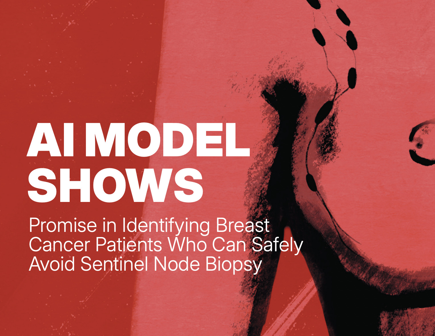
An AI model has been trained using mammograms to identify breast cancer patients who could be spared from unnecessary lymph node biopsy procedures. The study, published in NPJ Digital Medicine, 10 July, found that the introduction of the AI model could allow over 40% of current axillary surgical procedures to be avoided.
“Our findings suggest that routine mammograms, particularly full-breast images, can enhance preoperative nodal status prediction. The approach could be easily implemented as a routine diagnostic procedure preoperatively and avoid complications associated with lymph node biopsy,” explains corresponding author Lisa Rydén, who is Professor of Surgery at Lund University, Sweden.
Currently, all breast cancer patients (with a few exceptions) are recommended to undergo sentinel node biopsy. Sentinel node biopsy is a surgical procedure that uses a radiotracer or blue dye to identify the first in a chain of lymph nodes in the axilla where lymphatic fluid from the tumour drains. The rationale behind the procedure (performed during breast surgery) is that if the first lymph node is cancer-free, subsequent lymph nodes will also be cancer-free, thereby avoiding the need for axillary clearance (where all lymph nodes are removed), which leads to complications such as shoulder problems and lymphoedema (swelling of the arm). “But even sentinel node biopsy comes with side effects which are similar to those experienced after axillary clearance, but at a lower rate. And for surgeons, operating times would be shorter if they didn’t need to use tracers,” Rydén tells CancerWorld.
The spread of breast cancer to the axilla affects approximately one in five breast cancer patients, with the remainder having no trace of cancer in the lymph nodes and therefore deriving no therapeutic benefit from the procedure. Although there has been a move toward de-escalation of axillary surgery, reliable non-invasive methods for accurately assessing the risk of by axillary lymph node metastasis have been lacking.
Rydén and colleagues set out to devise an AI decision-support tool that could be used to predict the likelihood of axillary lymph node metastasis and identify patients who could safely forgo sentinel node biopsy. “We developed our algorithm in three steps,” explains Daqu Zhang, the first author of the study. “Firstly, the AI model went through tens of thousands of mammograms to learn their basic structure, such as edges, texture, and shapes. The AI model was then trained to find specific clues for cancer, such as the boundaries of tumours. And finally, it was given a ‘holistic mindset’ by including other important patient information, like age and tumour type, in order to more accurately predict the risk of metastasis.”
For the study, 1,265 women with clinically node negative (CNO) T1-T2 invasive breast tumours from three Swedish institutions who underwent surgery as a primary treatment between 2009 and 2017 were retrospectively included in the supervised learning cohort. Of these, 1,039 women (from sites 1 and 2) were included in the development set, 123 (from site 2) in the independent test set, and 103 (from site 3) in the external test set.
An innovative aspect of the study was the introduction of ‘Transformer neck’, an AI technique that allows the model to identify information regarding the risk of metastasis from the whole mammogram and not just the part containing the tumour.
Results showed that, in comparison to models using only clinical variables, incorporating full-breast mammograms with preoperative clinical variables improved the receiver operating characteristic (ROC) area under the curve (AUC) from 0.690 to 0.774. Put in context, AUC is a measure used to evaluate the capability to detect disease of interest (in this case, nodal metastasis) and the ability to correctly exclude healthy patients. “A value of 1.0 is considered perfect, while a value of > 0.7 is considered good, and > 0.8 very good,” explains Rydén.
Furthermore, if the model had been used in a breast cancer population with the same characteristics, the team would have been able to reduce the number of sentinel lymph node biopsy procedures undertaken by 41.7% (13.0–62.6%).
“These results highlight the great value of routine mammograms in staging LNM [lymph node metastasis] before surgery and aiding in preoperative patient stratification for axillary management by increasing the SLNB [sentinel node biopsy] reduction rate from 27% to 42%,” conclude the authors. “The innovative design of the Transformer neck, leveraging the attention mechanism, enhanced global feature extraction by emphasising important features in high-resolution, full-breast mammograms.”
Limitations of the study, write the authors, include the lack of diverse ethnic groups and that the external test set was not representative of clinical predictors for nodal status (like tumour size).
Further external validation is currently being undertaken with international collaborators. The investigators hope to add other data sources to the model, including gene expression data and images depicting histopathological sections of the breast tumour.
In the future, the authors believe that the AI algorithm could be used during routine mammography screening to assess the risk of lymph node metastasis. “Our article focuses on the spread to the lymph nodes, but in ongoing international studies, the image pattern could also be used to predict the prognosis,” says Rydén.
Independent Expert Comment
Douglas Flora, an oncologist and the Executive Medical Director of Oncology Services at St. Elizabeth Healthcare Cancer Center, Edgewood, Kentucky, with a special interest in the future of AI in cancer care, discusses the implications of the study with CancerWorld.

Could you comment on the overall significance of the study and what you see as the clinical importance of the findings?
The study demonstrates a significant advance in non-invasive risk stratification for early-stage, clinically node-negative (cN0) breast cancer patients. Its main contribution is successfully leveraging routine full-breast digital mammograms—a widely available and low-cost imaging modality—as a rich source of predictive information for axillary lymph node metastasis (LNM). The use of advanced deep learning (DL) techniques, specifically the Vision Transformer architecture, was crucial. This model improved the ability to predict LNM by recognising subtle patterns across the entire image (the ‘full-breast’) rather than just focusing on the tumour region of interest (ROI). This technique boosted performance significantly over models using only clinical variables, fundamentally elevating the mammogram from a detection tool to a powerful prognostic indicator.
From the clinical perspective, the most critical implication is the potential for de-escalation of axillary surgery. Reduction in Sentinel Lymph Node Biopsy (SLNB): the combined model (PreopClinic + FullMammo), operating under the stringent clinical constraint of maintaining a sensitivity of 90%, suggested a SLNB reduction rate of 41.7%. This means the tool could safely identify a large proportion of patients who might potentially omit SLNB, reducing the burden of postoperative complications without compromising oncologic safety.
The DL model proved as informative as key postoperative pathological indicators, such as pathological tumour size and multifocality. By accurately estimating risk factors before surgery, the model enables truly preoperative risk stratification, which is essential for informed surgical planning and patient counselling.
What do you see as the unanswered questions arising from the study?
The study itself highlights several important limitations and unknowns:
- Site-Dependent Variability: The model’s added predictive value varied considerably between the three Swedish institutions (Site 1, Site 2, and Site 3). The underlying causes for this are not yet identified but may relate to temporal shifts in diagnosis (e.g., earlier detection over time), differences in mammography equipment/vendors, or subtle variations in clinical workup protocols.
- External Validation Representativeness: The external test set (Site 3) was statistically unrepresentative of the development cohort in terms of key predictors like tumour size and LNM prevalence, limiting the generalisability of the external validation attempt.
- True Preoperative Data: The study used postoperative pathological assessment data (histological grade, type, molecular profile) as ‘preoperative predictors,’ which, while often available from core needle biopsy, is an assumption. The accuracy using only actual core needle biopsy measurements in a real-time clinical setting needs confirmation.
- Biological Basis of Global Features: While the Transformer identified important tumour and peri-tumour regions, the exact biological or microenvironmental changes in the breast tissue corresponding to the ‘global imaging patterns’ associated with LNM and LVI remain biologically uninterpreted.
What further research would you like to see undertaken?
I’d like to see the following research:
- Prospective Multicenter Validation Trial: A large-scale, prospective clinical trial is necessary to definitively validate the reported SLNB reduction rate. This trial should explicitly include diverse patient cohorts, different geographical regions, and a broad range of mammography equipment vendors to robustly address observed site-dependent variability.
- Mechanistic Feature Analysis (Biomarker Correlation): Research should employ advanced explainable AI (XAI) to map the AI-identified global imaging patterns to known biological phenomena. This involves correlating the image features with gene expression, immune cell infiltration, or other markers of the tumour microenvironment to understand why the model is predictive.
- Multimodal Data Fusion: Future models should incorporate the full-breast mammography features with other readily available preoperative data, specifically core-needle biopsy data (histology, molecular) and potentially sonographic features, to create a truly multimodal, high-performing prognostic tool.
- Long-Term Outcome Analysis: Validation must extend beyond immediate nodal status prediction to long-term oncologic outcomes, such as local recurrence and disease-free survival, for patients who safely omitted SLNB based on AI scores.
What would you say to cancer patients concerned about the findings of this study?
This is very encouraging research that points potentially toward a smarter, gentler way to manage breast cancer in future. For many women with early-stage breast cancer, the SLNB is a necessary step, but for about two-thirds the results are negative, meaning surgery was solely for staging. This study shows that a new AI tool can now look at standard mammograms and spot subtle, previously invisible patterns that strongly predict whether lymph nodes are cancer-free.
The key takeaway is that this technology suggested it could safely identify over 40% of patients who might someday be able to skip the SLNB entirely. This means we are moving closer to a future where we can personalise treatment even further, potentially avoiding unnecessary surgery and reducing risk of side effects,without compromising safety. While this tool is still in the research phase (needing testing in large-scale clinical trials before it becomes a standard part of care), it represents a significant step forward in making breast cancer treatment more precise and less invasive.
How is AI likely to be incorporated into oncology in the future?
AI integration into oncology is expected to proceed along three main tracks:
Operational Efficiency and Access (The System Optimiser): AI will enhance the functioning of the entire healthcare system. This includes optimising patient flow, managing resource allocation (e.g., scheduling biopsies, OR time), automating administrative tasks, and identifying and mitigating structural barriers to timely care. Ultimately, AI will drive overall efficiency and improve equitable access to high-quality treatment.
Augmented Diagnostics and Triage (The Co-Pilot): AI will become an essential partner for clinicians. In imaging (radiology) and tissue analysis (pathology), AI models will autonomously screen studies, flag subtle anomalies, and quantify disease features (e.g., predicting tumour size from mammograms, as seen in this study, or rapidly grading tumour aggressiveness). This will increase diagnostic speed, reduce false negatives, and allow human specialists to focus attention on the most complex cases.
Personalised Prognostication and Treatment Selection (The Navigator): AI will move beyond diagnosis to become a predictive engine. It will integrate complex data streams—genomics, clinical variables, and radiomics (image-derived features)—to create highly individualised risk scores. This will enable clinicians to select optimal treatments (e.g., which chemotherapy, which targeted agent, or, as in this study, whether surgery can be safely omitted) and predict the likelihood of treatment response and toxicity.