
Diffusion tensor imaging (DTI), a form of MRI, is capable of detecting pre-malignant lesions in both mouse and human pancreatic specimens. The ‘proof of concept’ study, published in Investigative Radiology, 13 December 2024, opens the way to use DTI for early diagnosis in people at risk of pancreatic cancer, and to understand more about carcinogenesis.
“This study represents a first step towards the early detection of pancreatic cancer with magnetic resonance imaging, even before the cancer develops,” says Carlos Bilreiro, the first author from the Radiology Department at the Champalimaud Clinical Centre in Lisbon. “We conclude that pancreatic intraepithelial neoplasia (PanIN) can be detected and characterised with DTI in mice, and that the findings are generalisable to specimens taken from human pancreas.”
Because symptoms of pancreatic cancer develop late in the course of the disease, many patients are diagnosed at advanced stages, too late to be eligible for pancreatic resection surgery, which is the only potentially curative therapeutic option. When patients are diagnosed in early stages, however, their outlook is much improved, with five-year survival rates of 25% to 50%.
Pancreatic ductal adenocarcinoma (PDAC) accounts for more than 90% of pancreatic cancers, and has several known precursor lesions (lesions that evolve into cancer). The most frequent precursor is pancreatic intraepithelial neoplasia (PanIN) – a lesion known to foreshadow 85% to 90% of PDACs. Noam Shemesh and Carlos Bilreiro, both from the Champalimaud Foundation, Lisbon, suggested that identifying PanIN could provide opportunities for early diagnosis of PDAC. However, until now the difficulty has been that PanIN lesions could not be diagnosed with current imaging modalities, including MRI.
Together, Shemesh and Bilreiro hypothesised that high-resolution DTI, with high sensitivity towards microarchitecture, would be capable of detecting and characterising microstructural changes in PanIN and early PDAC. “DTI is an MRI technique that relies on the diffusion of water molecules inside tissues. Because water molecules diffuse within the cells and experience interactions with cell walls and other microscopic objects, they serve as an endogenous tracer for tissue microstructure,” explains Shemesh, the corresponding author, who heads the Preclinical MRI lab at Champalimaud Research. Clinically, DTI is most commonly used for tractography in neuroimaging and to characterise neurological diseases.
For the study, the team used powerful preclinical MRI scanners (designed for use with small rodents and ex-vivo tissue specimens) that are considerably more powerful than the MRI scanners used clinically for imaging patients (16.4 Tesla vs 1.5 Tesla or 3 Tesla).
First, they focused on imaging two genetically engineered mouse models of PanIN and pancreatic cancer – the KC model and KPC model – on a monthly basis, until abnormalities were observed. KC mice develop PanIN over time, and may progress to pancreatic cancer in advanced stages; while the KPC models shows a more aggressive phenotype, resulting in earlier development of both PanIN and pancreatic cancer. “This part of the study was aimed at determining the time of onset of disease in both lines of transgenic mice, so that we could plan our experiments. Having the two models and controls allowed us to explore different degrees of disease burden and severity,” Bilreiro tells Cancerworld.
Once the team had identified gross changes in the structure of the pancreas with anatomical scans (signal heterogeneity and cystic changes), they went on to scan the changes in greater detail using DTI, looking for ‘hot spots’ that might suggest pathology. Next, after sacrificing the mice, the mouse pancreases underwent histopathological diagnosis that allowed the actual disease status (presence of PanIN, PDAC or normal pancreas) to be established for each animal. Healthy mice (with no genetic tendency to develop PanIN and PDAC) were used as controls to explore sensitivity and specificity.
Diffusion tensor imaging of pre-cancerous lesions in the pancreas
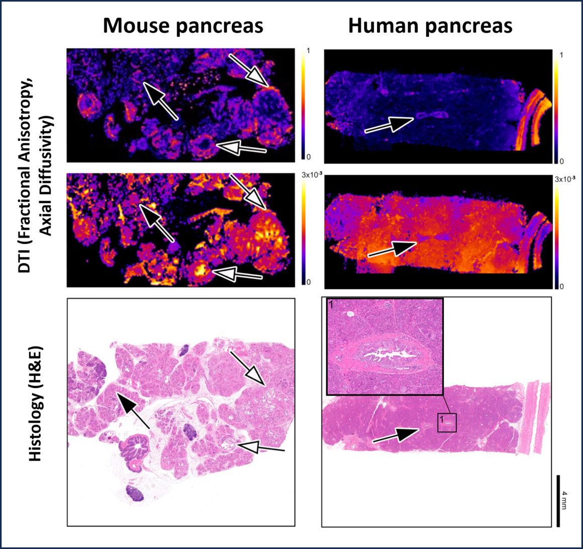
Source: Adapted from C Bilreiro et al. (2024) Pancreatic Intraepithelial Neoplasia Revealed by Diffusion-Tensor MRI. Investigative Radiology, courtesy of the investigators
Results showed correlation between the DTI-identified ‘hot spots’ and the histopathological diagnosis (using control mice for the comparison), with up to 100% sensitivity and specificity.
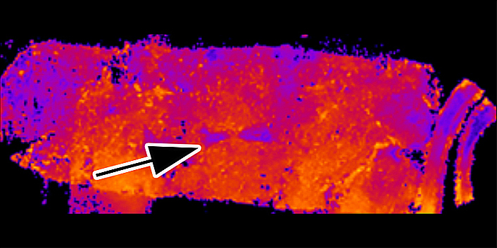
Finally, the team imaged biobank samples taken from five patients who underwent surgery for PDAC, with specimens sliced and placed inside a 10-mm MRI tube. “The histology and pathology of the samples showed that DTI was also efficient and effective for detecting human lesions,” says Shemesh.
The team view their study as ‘proof of concept’. “We have found a contrast approach that is relevant for diagnosing PanIN in humans and wanted to share these important findings early with other investigators,” Shemesh tells Cancerworld.
DTI technology, they believe, could ultimately be used as a method to screen for PanIN/ early signs of pancreatic cancer in people at high risk of developing the disease. “If we could identify PanIN early, we might be able to intervene before the cancer is established and improve prognosis,” says Bilreiro. DTI technology, he adds, is also likely to play a role in helping to understand the tumorigenesis of PanIN and its evolution to PDAC, an area that is currently little understood.
Although current clinical MRI machines will require technical adaptation (e.g. in image processing) to undertake DTI of the pancreas, the investigators are confident that these changes are relatively straightforward to make, and within the competence of most radiologists.
Next, Shemesh and Bilreiro hope to evaluate animals from the same transgenic line at different stages of disease progression to explore whether DTI is capable of staging PanIN and PDAC. Ultimately, they hope to validate the DTI imaging technology in patients prior to undergoing surgery for pancreatic cancer.